III. TBI-TECHNIQUE |
Gnotobiotic conditions.
Sources.
Beam incidence.
Beam quality.
I = D20 / D10 ( f+20 / f+10 )2
where D20/D10 represents the quotient of dose. Under TBI conditions, these doses have to be measured at 20 and 10 cm depth respectively in a phantom of at least 30 x 30 x 30 cm3. f is the source to phantom surface distance. Any beam quality can be used in AP-PA TBI. Bilateral TBI requires high energies.
Technical and geometrical data.
The more usual option is to provide a large source-patient distance (ranging from 3 to 6 m), and the use of the diagonal of the field for the positioning of the patient. It must be remembered, that the flattened part of the beam is always much smaller than that indicated by the optical alignment. There are techniques which utilize smaller treatment distances by using multiple parallel and adjacent beams, or moving beams or patient translation; see figure 1[3,6]
Patient position.
Treatment verification.
The most usual procedures for verification are the use of portal imaging methods to control the position of the shielding or alignment of detectors for in vivo dosimetry.
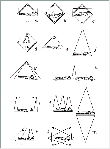
Figure 1.- TBI, single source: techniques using horizontal photon beams for bilateral (a,b) and for AP-PA irradiation in decubitus and standing position (c,d) and techniques using vertical photon beams for AP-PA irradiation with a large aperture 60Co-irradiator (e), large distance -two floors- (f), moving beam by head rotation (g), by patient translation (h), and horizontal source scan (i). Multiple beam TBI: techniques using adjacent fields (j) or direct and oblique fields (k). Multiple source TBI techniques (l,m).
RECOMMENDATIONS - STANDARD PARAMETERS OF TBI
|
Dosimetric measurements performed in antropomorphic phantoms are desirable.