Absolute dose
Dosemeter, detector and cable.
Attention should be paid to the response of the chosen detector and dosemeter, which must be as independent as possible, to the energy spectrum at TBI conditions. In these conditions the length of the cable irradiated is greater than in standard distances. The influence of this irradiation may be important in the measurement, when a small volume ionization chamber is used, because the current generated in the cable and stem may be comparable to the current produced in the air cavity[31,32].
Dose monitor and absorbed dose calibration.
Accuracy.
Relative dose
Beam fluence profile.
Beam uniformity decreases as treatment distance increases. The useful field length or beam diagonal and the flattened (±10%) part of the beam, have to be determined by measuring the beam profiles. Larger variations of photon beam fluence across the field have to be considered for dose calculations at different points on the patient.
When measuring the beam fluence profile, the main interest is the primary radiation. Therefore special care has to be taken with the choice of phantom size to avoid smearing.
Quasi primary radiation.
The inverse square law does not apply with the large distances used in TBI due to wall and floor scatter contributions. Empirical study of this law is of interest not only because of the treatment distance used but also, and mainly, because not all beam entrance points are at the same distance from the source[8].
Attenuation and scatter, phantom geometry.
The special conditions used in TBI imply different scatter conditions from normal treatment. In this case the contribution of walls and floor may not be negligible.
The different volumes of various parts of the patient's body create scatter conditions dissimilar from that of calibration[34-36]. Therefore relative data are needed to quantify this effect.
The finite volume of patients needs systematic measurements.
Depth dose, depth dose homogeneity.
In order to determine the dose at different points of the body, a comprehensive set of data has to be collected to permit the calculation of the required dose. The classical procedure is the use of Percentage Depth Dose (PDD) curves (TPR curves may be used as well) normalized at the reference depth, and measured under TBI conditions. An advisable extra set of data could include, in addition to the central axis curve, curves which are parallel to this, which may also be taken under different conditions since depth dose depends on patient thickness, beam quality, source-skin distance, field size, distance to central axis and the presence of different scattering materials.
The ratio of the maximum depth dose relative to the minimum one (for opposing beams it is usually the midplane dose or the skin dose) describes the depth dose homogeneity.
Tissue heterogeneity, lung corrections.
By using the water density as a reference for the whole body, errors can be introduced in areas such as lungs. A useful first approach is the tissue equivalent concept. This considers the density of the traversed tissues and takes the anatomical information from radiographs or CT taken under TBI conditions. However, this correction only takes into account the primary radiation and neglects scattered radiation. More precise corrections require more complex measurements and planning algorithms.
Attention should be paid to the fact that lung density varies with the age of the patient, the position (the density of the lung closer to the couch is always higher than the density of the other side), and zone (upper or lower)[30]. Nevertheless, lung correction factors depend not only on density but also on body size and thickness, lung size and thickness, thorax wall thickness and shielding size and thickness.
Dose modifier, partial shielding, bolus, compensators.
One of the main modifiers is the shielding for the lungs. Dose contributions due to secondary electrons may be important if the distance from the shield to the skin is small. This effect is generally avoided but in some cases it may be useful to prevent under irradiation of the skin. At the same time, as this distance increases or if the modifier is placed in an unstable place, the possibility of misalignment rises.
Other dose modifiers include the use of bolus for missing tissue compensation. This compensation is essential in bilateral irradiation if midline dose homogeneity is to be achieved. It should be recognised that a compensator placed close to the skin will behave in a different way than the one remotely positioned. The former contributes with both attenuation and scatter while the latter produces mainly attenuation.
Surface dose, scatter screen/spoiler.
If the skin is to be treated as part of the target volume, an estimation of the dose at the skin, both entrance and exit, is needed. This problem becomes harder as the beam energy increases. An appropriate thickness of suitable material placed close to the skin will ensure improved electronic equilibrium at the patient's skin. For this reason beam spoilers are typically used. In these cases the "spoiler to skin" distance is generally different along the patient due to the patient's contour. These different distances may produce different contributions along the body[30]. Blankets, pyjamas, coverlets or sheets may be used to increase the skin dose[37].
In-vivo dosimetry
In normal TBI practice, many centres do not utilize in-vivo dosimetry. Other centres, however, base their plannings on in vivo measurements[38]. In-vivo dosimetry needs special equipment and some considerable effort to reach sufficient accuracy.
Detectors.
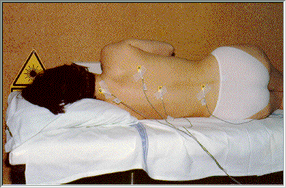
To check the agreement between the prescribed and the delivered dose an "in vivo" dosimetry system (TLD or semiconductors) is commonly used during every treatment as well as for treatment planning, conformation or verification and for dose monitoring.
Although the accuracy of TLD detectors may be high and depend only on a few parameters the results are delayed. On the other hand, semiconductor probes work on line, although their response depends on parameters such as energy, temperature, radiation damage, dose rate and directionality[30,39-46].
Calibration.
RECOMMENDATIONS - TBI DOSIMETRY
- As a general rule, all dose measurements have to be performed under TBI conditions, in water and as close to the real treatment situation as possible. Water will be used as a reference material. If a plastic substance is used, the measurement will be related to water. All relative measurements have to be related to the calibration point.
- The dosimeters must be calibrated periodically against the national standards and the recommendations of the recognized dosimetric protocol (NACP, IAEA, SEFM, IPSM, etc.) must be followed[47-50].
- An estimation of the effect of the irradiation of cables and stems should be made.
- The absolute dose calibration must be done using a phantom with at least 30 x 30 x 20 cm3. The depth of the measurement point should be larger than the depth of maximum dose, and preferably 10 cm which corresponds to the average midthickness of a patient. The dose unit is Gy.
- The dose monitor should be calibrated against dose measurement at the centre of the square calibration phantom.
- The estimated accuracy required should be stated. The accuracy should be as good as possible, preferably below ±5% (95% confidence level).
- At least 4 beam profiles, lateral and diagonal, should be measured.
- Beam flattening must be checked, and corrected, if necessary by using additional flattening filters.
- Free in air measurements should be avoided.
- Measurements of beam profiles should be made with a build-up cap of 10 cm water equivalent thickness and related to the reference dose at calibration conditions.
- Wall and floor scatter contributions must be considered.
- The inverse square law must be verified within the range of distances foreseen in TBI treatments. If the differences are higher than 2%, an empirical curve should be determined.
- The lack of backscatter due to finite thickness should also be taken into account.
- To consider the different volumes of parts of the patient's body, measurements under total and partial scattering conditions should be made.
- In order to describe the influence on depth dose, measurements should be performed with phantoms of different sizes and thicknesses, at different source-skin distances. Dose data, in addition to the central axis curve, should be measured, because differences may not be negligible.
- Lung dose correction factors have to be determined for the thickness of the thorax wall, lung density as well as the shielding thickness and size.
- Partial shielding of the lung must be used in every session in order to keep the same set of TBI parameters during all fractions.
- In order to achieve a better dose homogeneity, an appropriate thickness of a suitable material (spoiler) may be placed in front of the patient to ensure electronic equilibrium at the patient's skin, when necessary.
- An in-vivo dosimetry system should be available to check the prescribed dose or to monitor the machine.
- Calibration of detectors for in-vivo dosimetry must be performed at suitable phantoms and under TBI treatment conditions in order to consider all relevant factors.
|
[ Back ]
[ Index ]
[ Next ]


